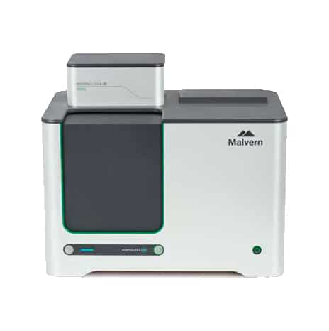
Morphologi 4-ID
Malvern panalytical
Morphologi 4-ID delivers detailed component-specific morphological descriptions of particulate blends through Morphologically-Directed Raman Spectroscopy (MDRS).
Rapid, automated, component-specific particle characterization in a single, integrated platform with one-year subscription to Wiley’s KnowItAll® Raman Identification Pro package
Advantages
The Morphologi 4-ID offers an exclusive capability, combining all the benefits of automated static imaging delivered by the Morphologi 4 with chemical identification of individual particles by Raman spectroscopy in a single measurement.
- Patented MDRS capability delivers representative component-specific particle size and shape data, delivering full characterization of your sample
- All the functionality of the Morphologi 4 combined with a dedicated Raman platform for both physical and chemical particle characterization in a single, automated measurement
- Automatically measures Raman spectra for hundreds or thousands of particles, saving valuable analyst time
- Intuitive software ensures suitability for experienced and non-experienced spectroscopists alike
- Easy correlation of morphological properties with chemical information provides the most comprehensive understanding of your sample
- 21 CFR Part 11 software option ensures regulatory compliance
- Conforms to standards: ISO 9276-6 and ISO 13221-1
The Morphologi 4-ID measurement procedure can be split into five sections:
Sample preparation
Spatial separation of individual particles and agglomerates is critical to robust results. The integrated dry powder disperser makes preparing dry powder samples easy and reproducible. The applied dispersion energy can be precisely controlled, enabling the measurement process to be optimized for a range of material types. Accessories that fit directly in to the Morphologi 4-ID’s automated stage are available for preparing suspended or filtered samples.
Image capture
The instrument captures images of individual particles by scanning the sample underneath the microscope optics. The Morphologi 4-ID can illuminate the sample from below or above, whilst accurately controlling the light levels.
Image processing
Use of either the automated ‘Sharp Edge’ segmentation analysis or the manually-controlled thresholding enables the detection of particles and the calculation of a range of morphological parameters for each.
MDRS
Components and particles of interest are targeted for Raman analysis. Particles can be selectively targeted based on their morphology, or particles representative of the whole sample can be targeted objectively by the Morphologi software.
Result generation
Advanced graphing and data classification options in the software ensure that extracting the relevant data from the measurement is straightforward, via an intuitive visual interface. Correlation scores to a reference library are calculated for each Raman spectrum acquired, enabling particles to be classified based on their chemistry. The morphological data associated with the particles in each chemical class is used to generate size and shape distributions, delivering component-specific morphological information. Individually-stored grayscale images for each particle are linked to their Raman spectra and provide qualitative verification of the quantitative results.
| Technology | Static automated imaging |
|---|---|
| Particle size | 0.5 μm – 1300 μm (upper limit may be extended for some applications*) |
| Particle properties measured | Size, shape, transparency, count, location |
| Particle size parameters | Circle equivalent (CE) diameter, length, width, perimeter, area, maximum distance, sphere equivalent (SE) volume, fiber total length, fiber width |
| Particle shape parameters | Aspect ratio, circularity, convexity, elongation, high sensitivity (HS) circularity, solidity, fiber elongation, fiber straightness |
| Particle transparency parameters | Intensity mean, intensity standard deviation |
| Integrated Sample Dispersion Unit | For fully automated dispersion and measurement of dry powders. Manual or SOP control of dispersion pressure, injection time and settling time |
| Illumination | White light LED: brightfield, diascopic and episcopic; darkfield, episcopic |
| Detector | 18 MP; 4912 x 3684 pixel color CMOS array; pixel size 1.25 μm x 1.25 μm |
| Optical system | Nikon CFI 60 brightfield / darkfield system |
| Lens (and particle size range) | 2.5x: 8.5 µm – 1300 µm (nominal) 5x: 4.5 µm – 520 µm (nominal) 10x: 2.5 µm – 260 µm (nominal) 20x: 1.5 µm – 130 µm (nominal) 50x: 0.5 µm – 50 µm (nominal) |
- Building materials
- Energy storage/batteries
- Forensics
- Mining and minerals
- Pharmaceutical development
